
Laparoscopic & robotic diaphragm plication
Stabilizing a weak diaphragm to create more room to breathe.
A paralyzed diaphragm does not always require surgery. When one side remains elevated and causes activity-limiting shortness of breath, diaphragm plication may improve breathing mechanics. Dr. Paresh Mane offers laparoscopic and robotic-assisted approaches for selected patients after a careful evaluation.
Request an evaluationWhat is a paralyzed diaphragm?
The diaphragm is the broad muscle separating the chest from the abdomen. Each half is controlled by a phrenic nerve. During a normal breath, the diaphragm contracts and moves downward so the lungs have room to expand.
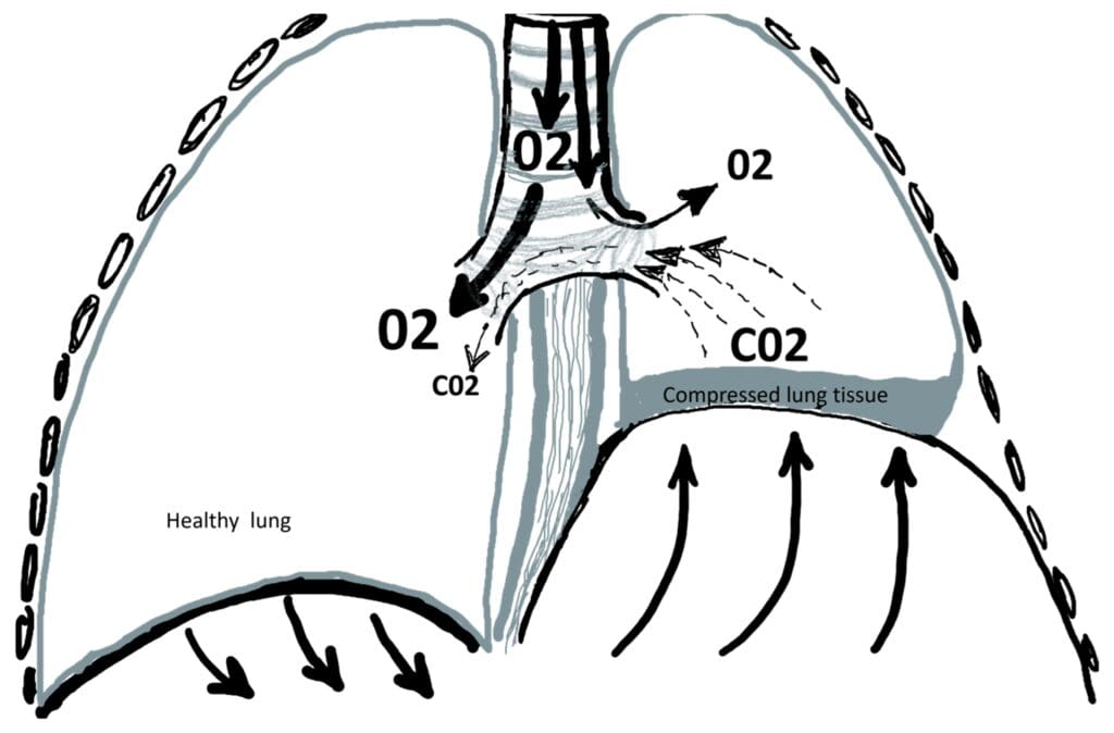
Phrenic-nerve injury or compression may follow heart, chest or neck surgery; trauma; cervical spine disease; a tumor; or a neurological or neuromuscular disorder. Sometimes no cause is found. An elevated hemidiaphragm on an X-ray does not by itself prove paralysis, so the cause and actual motion need to be assessed. [2]
One-sided paralysis may cause shortness of breath with exertion, difficulty breathing when lying flat, reduced exercise tolerance, fatigue or sleep problems. Some people have no symptoms. Bilateral paralysis is a different, potentially more serious problem and requires respiratory and sometimes neurological evaluation rather than automatically proceeding to plication.
How is diaphragm paralysis evaluated?
The goal is to confirm abnormal diaphragm motion, identify a possible cause and determine how much it contributes to breathing symptoms. Testing is individualized and may include:
- Chest X-ray: identifies an elevated side of the diaphragm.
- Dynamic ultrasound or fluoroscopic sniff test: shows how the diaphragm moves during breathing and a quick sniff.
- Pulmonary-function tests: measure breathing capacity; comparing upright and supine results can be informative.
- CT imaging: evaluates the chest, diaphragm and possible causes of phrenic-nerve compression.
- Additional testing: sleep, cardiac, neurological or phrenic-nerve testing may be appropriate when the diagnosis remains uncertain. [1]
Other causes of shortness of breath—such as lung disease, heart disease, anemia, deconditioning or obesity—also need consideration. Plication is most likely to help when symptoms and objective diaphragm dysfunction fit together.
Who may benefit from diaphragm plication?
Plication is generally considered for an adult with persistent, lifestyle-limiting symptoms attributable to one weak, paralyzed or eventrated hemidiaphragm. The decision is based on symptom severity, diaphragm motion, lung-function results, timing and the likelihood that another condition is driving the breathlessness.
| Situation | What may be discussed |
|---|---|
| Mild or no symptoms | Observation and treatment of contributing conditions; surgery is usually unnecessary. |
| Recent phrenic-nerve injury | A period of observation may be appropriate because nerve function can recover. Timing is individualized rather than based on one rule. |
| Persistent, limiting symptoms | Plication may be considered when testing confirms that the elevated diaphragm is an important cause. [1] |
| Bilateral dysfunction | A separate respiratory and neuromuscular assessment is needed; treatment may differ from one-sided paralysis. |
The purpose of surgery
What does diaphragm plication do?
Plication places the loose, elevated diaphragm in a flatter and lower position. The surgeon creates folds and secures them with strong sutures. This limits paradoxical upward movement, reduces compression of the lung and leaves more room for the lung to expand. [3]
- Access the diaphragm. Under general anesthesia, the surgeon reaches the diaphragm through small abdominal or chest incisions, depending on the chosen approach.
- Set the position. The elevated muscle is brought into a lower, appropriately tensioned position while the lung and abdominal organs are protected.
- Secure the repair. Rows of durable sutures create folds that hold the diaphragm flatter. A chest tube is used only when the operative findings and route make one necessary.
Small-incision abdominal access
Laparoscopic and robotic-assisted plication
Both approaches can reach the underside of the diaphragm through several small abdominal ports. Carbon dioxide creates working space, a camera provides the view and the surgeon places sutures to tighten the diaphragm. This avoids a rib-spreading incision, but it remains major surgery and may need to change to an open approach for safety.
Laparoscopic
The surgeon operates a camera and long instruments directly through abdominal ports. This provides a broad view of the abdominal surface of the diaphragm and nearby organs.
Robotic-assisted
The operation follows the same plication principle. From a console, the surgeon controls wristed instruments and a magnified 3D camera, which can help with precise suturing in a confined area. The robot does not operate independently.
Approach selected for you
The side involved, body shape, previous chest or abdominal surgery, adhesions, other procedures and surgeon experience all influence whether abdominal, chest-based or open access is safest.
A laparoscopic or robotic abdominal route may be useful when avoiding intercostal chest incisions is desirable. A thoracoscopic or robotic chest approach may instead offer the best access in another patient. No high-quality evidence shows that one minimally invasive route is best for everyone. [4]
Recovery and risks
Hospital care: the length of stay depends on breathing, pain control, mobility and whether a chest tube or additional treatment is needed. Minimally invasive reports often describe short stays, but your operation and recovery plan are individual.
At home: early walking and deep breathing are usually encouraged. Soreness and fatigue improve gradually, and breathing benefit may unfold over weeks to months. Follow the hospital and surgical teams’ instructions for medications, wound care, lifting, driving and return to work. [7]
Risks to discuss include:
- Bleeding, infection, pain and reactions to anesthesia.
- Pneumonia, atelectasis or other breathing problems.
- Air or fluid around the lung, an air leak or the need for a chest tube.
- Injury to the lung or nearby abdominal organs such as the liver, spleen, stomach or bowel.
- Blood clots, heart-rhythm problems or ileus.
- Persistent symptoms, recurrent elevation or failure of the plication.
- Conversion to a larger abdominal or chest incision when needed for safety.
Ask what benefit is realistic for your symptoms, which approach is proposed, why that route is recommended and how complications would be managed.
Common questions
Is robotic surgery always better than laparoscopy?
No. Robotic instruments can make suturing easier in some situations, while conventional laparoscopy may be equally appropriate in others. Current evidence does not prove one technique is superior for every patient.
Will plication make the diaphragm move normally again?
Usually not. Plication holds the weak side in a more useful position; it does not reconnect or repair the phrenic nerve.
Could a minimally invasive operation become open?
Yes. Scar tissue, anatomy, bleeding or another unexpected finding can make a larger incision the safest way to complete the operation.
Do I need to wait before considering surgery?
Recent nerve injuries can improve with time, so observation may be appropriate. Persistent severe symptoms may justify an earlier discussion. Your cause, duration, testing and daily limitations determine the timing.
New England Diaphragm Center
Discuss diaphragm plication with Dr. Mane
Contact Merrimack Thoracic & Esophageal Surgery for an evaluation or second opinion, including whether laparoscopic or robotic-assisted diaphragm plication may fit your situation.
Bring prior chest imaging, sniff-test or ultrasound results, pulmonary-function tests and records of chest, heart, neck or abdominal surgery. Ask the office how to transfer medical records securely.
Referral fax: (978) 346-3000
Sources and further reading
Original patient information based on academic clinical guidance and published research. Sources checked September 12, 2026. This page supports—but does not replace—an individual surgical assessment.
- Gilbert & Wei: Diaphragmatic plication—current evidence and techniques (2023)
- Dubé & Dres: Diaphragm dysfunction—diagnostic approaches and management strategies (2016)
- Baylor Medicine: Plication of the diaphragm
- Le et al.: Robotic diaphragm plication—functional and surgical outcomes (2023)
- Gritsiuta et al.: Systematic review of minimally invasive diaphragm plication (2022)
- Biswas et al.: Transabdominal robot-assisted diaphragmatic plication (2018)
- University of Utah Health: What to expect during diaphragm plication